Author: SAR-ADm26

  • Can cataracts come back after Surgery

    Can Migraines Cause Vision Problems That Need Neuro-Ophthalmology Care?

    “Doctor, I could see flashing lights for a few minutes, and then everything became normal. Was it just a migraine?”
    It’s a question eye specialists hear more often than you might think. Migraines are well known for causing severe headaches, but the headache isn’t always the first symptom. For many people, the first sign is a change in vision. A shimmering line appears out of nowhere. A blind spot makes reading difficult. Bright lights seem unusually intense. The episode passes, and life goes on.
    Most of the time, that’s exactly what happens. But not always.
    Knowing the difference is where neuro-ophthalmology care plays an important role.

    When a Migraine Changes What You See

    Vision isn’t created by the eyes alone. The eyes capture images, but the brain interprets them. During a migraine, temporary changes in brain activity can interrupt this process, leading to what doctors call a migraine aura.
    Not everyone experiences it in the same way.
    Some people notice flashing lights. Others describe zigzag patterns, blurred vision, or a patch where they simply can’t see. A few find that reading becomes difficult because the words seem to disappear from the centre of the page.

    When It May Not Be “Just Another Migraine”

    Our brains like familiar explanations.
    If you’ve had migraines before, it’s natural to assume every new visual disturbance is another episode. But medicine isn’t always that straightforward. A sudden change in vision can also signal problems involving the optic nerve, the nerves controlling eye movement, or even the brain itself. In other words, migraine causes vision problems, but so do several other neurological conditions.

    That’s why the context matters.

    Did the symptoms feel different this time?

    Was there double vision instead of flashing lights?

    These small details often guide the next step.

    The Eye Isn’t Always the Problem

    One of the biggest misconceptions is that every vision problem begins inside the eye.

    In reality, the eye is only the starting point. Once light enters the eye, the information travels through the optic nerve and several areas of the brain before you actually “see” anything.

    A disruption anywhere along this pathway can affect vision, even when the eye itself appears completely normal.

    This is exactly what neuro-ophthalmology care focuses on.

    Instead of looking only at the eye, a neuro-ophthalmologist evaluates how the eyes, optic nerves, and brain work together.

    Conditions That Can Mimic Migraine

    Several neurological conditions can initially resemble a migraine aura. Inflammation of the optic nerve may begin with blurred vision and discomfort while moving the eye.
    Raised pressure inside the skull can cause brief episodes where vision suddenly dims before returning to normal.
    Certain nerve disorders may present with double vision instead of blurred vision. In some cases, even a stroke or a transient ischaemic attack (TIA) can first appear as an unexpected visual disturbance.
    These are only a few examples of the many causes of vision problems that deserve careful evaluation rather than assumptions.

    When Should You See a Specialist?

    You should seek medical attention if you experience:
    • Sudden loss of vision in one or both eyes
    • Double vision
    • Visual symptoms lasting longer than an hour
    • A new type of visual disturbance you’ve never experienced before
    • Weakness, numbness, difficulty speaking, or loss of balance along with vision changes
    • A severe headache that feels very different from your usual migraines

    What Happens During a Neuro-Ophthalmology Consultation?

    A comprehensive eye examination is usually followed by tests that assess the optic nerve, peripheral vision, eye movements, or retinal health. If required, imaging studies such as an MRI or CT scan may also be recommended. Each test answers a different question, helping your doctor determine whether the symptoms are related to migraine or another neurological condition.

    Why an Accurate Diagnosis Matters

    If your symptoms are caused by migraine, identifying the pattern can help you manage future episodes with greater confidence. If they aren’t, early diagnosis can make all the difference.
    Many neurological conditions affecting vision respond well to treatment when recognised early. Waiting for repeated episodes or assuming every visual disturbance is another migraine may delay the care you actually need.
    If you’re experiencing recurring, unexplained, or unusual visual symptoms, consulting our neuro-ophthalmology specialist in Nagpur can help determine what’s really happening and guide the right course of treatment.
    At Sarakshi Netralaya, our approach to neuro-ophthalmology care goes beyond treating symptoms. We focus on understanding the connection between the eyes and the brain, using advanced diagnostics and clinical expertise to identify the real cause of your visual concerns, so that you receive the right treatment at the right time.

    Frequently Asked Questions (FAQs)

    Book an Appointment

    [wpforms id=”63″ title=”false”]
  • Can Migraines Cause Vision Problems That Need Neuro-Ophthalmology Care?

    Can Migraines Cause Vision Problems That Need Neuro-Ophthalmology Care?

    “Doctor, I could see flashing lights for a few minutes, and then everything became normal. Was it just a migraine?”
    It’s a question eye specialists hear more often than you might think. Migraines are well known for causing severe headaches, but the headache isn’t always the first symptom. For many people, the first sign is a change in vision. A shimmering line appears out of nowhere. A blind spot makes reading difficult. Bright lights seem unusually intense. The episode passes, and life goes on.
    Most of the time, that’s exactly what happens. But not always.
    Knowing the difference is where neuro-ophthalmology care plays an important role.

    When a Migraine Changes What You See

    Vision isn’t created by the eyes alone. The eyes capture images, but the brain interprets them. During a migraine, temporary changes in brain activity can interrupt this process, leading to what doctors call a migraine aura.
    Not everyone experiences it in the same way.
    Some people notice flashing lights. Others describe zigzag patterns, blurred vision, or a patch where they simply can’t see. A few find that reading becomes difficult because the words seem to disappear from the centre of the page.

    When It May Not Be “Just Another Migraine”

    Our brains like familiar explanations.
    If you’ve had migraines before, it’s natural to assume every new visual disturbance is another episode. But medicine isn’t always that straightforward. A sudden change in vision can also signal problems involving the optic nerve, the nerves controlling eye movement, or even the brain itself. In other words, migraine causes vision problems, but so do several other neurological conditions.

    That’s why the context matters.

    Did the symptoms feel different this time?

    Was there double vision instead of flashing lights?

    These small details often guide the next step.

    The Eye Isn’t Always the Problem

    One of the biggest misconceptions is that every vision problem begins inside the eye.

    In reality, the eye is only the starting point. Once light enters the eye, the information travels through the optic nerve and several areas of the brain before you actually “see” anything.

    A disruption anywhere along this pathway can affect vision, even when the eye itself appears completely normal.

    This is exactly what neuro-ophthalmology care focuses on.

    Instead of looking only at the eye, a neuro-ophthalmologist evaluates how the eyes, optic nerves, and brain work together.

    Conditions That Can Mimic Migraine

    Several neurological conditions can initially resemble a migraine aura. Inflammation of the optic nerve may begin with blurred vision and discomfort while moving the eye.
    Raised pressure inside the skull can cause brief episodes where vision suddenly dims before returning to normal.
    Certain nerve disorders may present with double vision instead of blurred vision. In some cases, even a stroke or a transient ischaemic attack (TIA) can first appear as an unexpected visual disturbance.
    These are only a few examples of the many causes of vision problems that deserve careful evaluation rather than assumptions.

    When Should You See a Specialist?

    You should seek medical attention if you experience:
    • Sudden loss of vision in one or both eyes
    • Double vision
    • Visual symptoms lasting longer than an hour
    • A new type of visual disturbance you’ve never experienced before
    • Weakness, numbness, difficulty speaking, or loss of balance along with vision changes
    • A severe headache that feels very different from your usual migraines

    What Happens During a Neuro-Ophthalmology Consultation?

    A comprehensive eye examination is usually followed by tests that assess the optic nerve, peripheral vision, eye movements, or retinal health. If required, imaging studies such as an MRI or CT scan may also be recommended. Each test answers a different question, helping your doctor determine whether the symptoms are related to migraine or another neurological condition.

    Why an Accurate Diagnosis Matters

    If your symptoms are caused by migraine, identifying the pattern can help you manage future episodes with greater confidence. If they aren’t, early diagnosis can make all the difference.
    Many neurological conditions affecting vision respond well to treatment when recognised early. Waiting for repeated episodes or assuming every visual disturbance is another migraine may delay the care you actually need.
    If you’re experiencing recurring, unexplained, or unusual visual symptoms, consulting our neuro-ophthalmology specialist in Nagpur can help determine what’s really happening and guide the right course of treatment.
    At Sarakshi Netralaya, our approach to neuro-ophthalmology care goes beyond treating symptoms. We focus on understanding the connection between the eyes and the brain, using advanced diagnostics and clinical expertise to identify the real cause of your visual concerns, so that you receive the right treatment at the right time.

    Frequently Asked Questions (FAQs)

    Book an Appointment

    [wpforms id=”63″ title=”false”]

  • Does Untreated Squint Affect Vision Long-Term? Risks, Complications & Treatment Options

    Does Untreated Squint Affect Vision Long-Term? Risks, Complications & Treatment Options

    Does Untreated Squint Affect Vision Long-Term?

    A squint, medically known as strabismus, is more than just an eye alignment issue. While many people assume it’s only a cosmetic concern, untreated squint can have lasting effects on vision, depth perception, and overall quality of life. In both children and adults, delayed treatment may lead to permanent visual complications that become harder to correct over time.
    Fortunately, advances in eye care have made squint treatment more effective than ever. With early diagnosis and personalized care from a squint treatment specialist, many patients can achieve better eye alignment and preserve their vision.
    Let’s explore how untreated squint affects the eyes, the potential long-term risks, and the treatment options available today.

    What Is a Squint?

    A squint occurs when the eyes do not look in the same direction at the same time. One eye focuses on an object, while the other may turn inward, outward, upward, or downward.
    The condition may be:
    • Present from birth (congenital)
    • Develop during childhood
    • Appear later in adulthood due to nerve, muscle, or medical conditions
    The brain receives two different images from the eyes, making it difficult to create a single clear picture.

    Why Does Squint Need Early Treatment?

    Normal vision depends on both eyes working together. When one eye is misaligned, the brain struggles to combine the images.
    In children, the brain often begins to ignore the image from the weaker eye to avoid double vision. While this adaptation may seem helpful, it can result in permanent vision loss if left untreated.
    Early crossed eye treatment helps restore proper eye coordination and supports healthy visual development.

    Can Untreated Squint Cause Permanent Vision Problems?

    Yes. One of the biggest untreated squint problems is that it can lead to long-term or even permanent visual impairment, especially when treatment is delayed during childhood.
    The severity depends on:
    • Age at onset
    • Type of squint
    • Degree of eye misalignment
    • How long the condition remains untreated
    Early intervention offers the best chance of preserving normal vision.

    Long-Term Problems Caused by Untreated Squint

    1. Lazy Eye (Amblyopia)

    One of the most common untreated squint problems is amblyopia, commonly called lazy eye. When the brain consistently ignores one eye, that eye gradually loses visual function.
    Without timely treatment:
    • Vision may never fully develop
    • Permanent reduction in vision can occur
    • Glasses alone may not restore eyesight

    2. Loss of Depth Perception

    Healthy vision requires both eyes to work together.
    Untreated squint can reduce or eliminate stereoscopic (3D) vision, making it difficult to judge distances accurately.
    This affects everyday activities such as:
    • Driving
    • Playing sports
    • Climbing stairs
    • Pouring liquids
    • Parking a vehicle
    Even simple daily tasks may become more challenging.

    3. Double Vision in Adults

    Unlike children, adults often cannot suppress the image from one eye.
    Instead, untreated squint may lead to:
    • Persistent double vision
    • Eye strain
    • Headaches
    • Difficulty reading
    • Problems concentrating
    Many adults seek crossed eye treatment because these symptoms interfere with work and daily life.

    4. Reduced Visual Comfort

    Misaligned eyes often work harder to focus.
    This can cause:
    • Eye fatigue
    • Blurred vision
    • Difficulty with prolonged screen use
    • Frequent headaches
    Treatment can significantly improve visual comfort.

    4. Reduced Visual Comfort

    Although squint primarily affects vision, it can also influence confidence and social interactions.
    Some individuals may experience:
    • Reduced self-esteem
    • Difficulty maintaining eye contact
    • Social anxiety
    • Emotional distress
    Correcting eye alignment often improves both vision and confidence.

    Is Squint Only a Childhood Condition?

    No.
    Although squint is more commonly diagnosed in children, adults can also develop it due to:
    • Diabetes
    • Stroke
    • Thyroid eye disease
    • Head injury
    • Nerve disorders
    • Previous eye surgery
    Adults experiencing newly developed squint should seek prompt evaluation, as it may indicate an underlying medical condition.

    How Is Squint Diagnosed?

    A squint treatment specialist performs a comprehensive eye examination that may include:
    • Visual acuity testing
    • Eye alignment assessment
    • Eye movement evaluation
    • Binocular vision testing
    • Refraction for glasses
    • Dilated retinal examination
    Advanced diagnostic equipment helps determine both the cause and severity of the condition.

    Modern Squint Treatment Options

    Treatment depends on the patient’s age, type of squint, and severity.

    Prescription Glasses

    Some forms of squint are caused by refractive errors. Corrective glasses may improve eye alignment in suitable patients.

    Vision Therapy

    Certain patients benefit from eye exercises that strengthen coordination between both eyes. Vision therapy is often recommended alongside other treatments.

    Patching Therapy

    For children with lazy eye, patching the stronger eye encourages the weaker eye to develop better vision.
    Early treatment provides the greatest benefit.

    Prism Glasses

    Special prism lenses help reduce double vision in selected adult patients.

    Botulinum Toxin (Botox)

    In some cases, carefully administered Botox injections temporarily relax eye muscles and improve alignment.

    Squint Surgery

    When non-surgical methods are insufficient, squint treatment may involve surgery to adjust the eye muscles.
    Modern crossed eye treatment surgery is:
    • Safe
    • Precise
    • Performed under anesthesia
    • Associated with faster recovery
    • Effective in improving eye alignment
    The goal is to restore better eye coordination and improve visual function whenever possible.

    When Should You See a Squint Treatment Specialist?

    Consult a squint treatment specialist if you notice:
    • Eyes that do not appear aligned
    • Frequent eye turning
    • Double vision
    • Head tilting while focusing
    • Closing one eye in bright sunlight
    • Poor depth perception
    • Persistent headaches related to vision
    Children should undergo routine eye examinations even if they do not complain of vision problems, as many cases of squint are first noticed during screening.

    Can Adults Benefit from Squint Treatment?

    Absolutely.
    Many adults assume treatment is no longer possible, but modern squint treatment can significantly improve:
    • Eye alignment
    • Double vision
    • Depth perception
    • Eye comfort
    • Cosmetic appearance
    • Quality of life
    Even long-standing squints can often be managed successfully after proper evaluation.

    Why Early Treatment Makes a Difference

    The earlier a squint is diagnosed, the better the chances of preventing permanent vision loss.
    Early treatment helps:
    • Preserve vision
    • Prevent lazy eye
    • Improve binocular vision
    • Restore eye coordination
    • Reduce future complications
    Delaying treatment increases the risk of irreversible visual impairment, particularly in young children.

    Why Choose an Experienced Squint Treatment Specialist?

    Managing squint requires careful evaluation of eye muscles, vision, and binocular function. An experienced squint treatment specialist can identify the underlying cause and recommend the most appropriate treatment—whether glasses, vision therapy, or surgery.
    Sarakshi Netralaya offers comprehensive diagnosis and advanced squint treatment using modern technology and individualized care plans for both children and adults. Patients looking for a trusted squint treatment hospital in Nagpur can benefit from expert evaluation and long-term follow-up.

    Frequently Asked Questions (FAQs)

    Conclusion

    Squint is far more than an alignment issue—it can significantly impact vision if left untreated. From lazy eye and reduced depth perception to double vision and long-term visual impairment, the consequences of delayed care can be serious. The good news is that modern squint treatment offers highly effective solutions for both children and adults.
    If you or your child has symptoms of a squint, don’t wait. Early consultation with a qualified squint treatment specialist can protect vision, improve eye coordination, and prevent lifelong complications. At Sarakshi Netralaya, expert care and advanced treatment options help patients achieve healthier vision and better quality of life.

    Book an Appointment

    [wpforms id=”63″ title=”false”]

  • Can Glaucoma Be Managed Without Surgery? Modern Treatment Options Explained

    Can Glaucoma Be Managed Without Surgery? Modern Treatment Options Explained

    Can Glaucoma Be Managed Without Surgery Using Modern Treatments?

    Glaucoma is often called the “silent thief of sight” because it usually develops without noticeable symptoms until significant vision loss has already occurred. According to the World Health Organization, glaucoma is one of the leading causes of irreversible blindness worldwide. The good news is that early diagnosis and timely glaucoma treatment can slow or even halt disease progression in many patients.
    A common question patients ask is, “Can glaucoma be managed without surgery?” The answer is yes, in many cases. Thanks to advances in ophthalmology, several non-surgical glaucoma modern treatment options are available that help control eye pressure, preserve vision, and delay or even eliminate the need for surgery.
    Let’s understand how modern glaucoma management works and when surgery becomes necessary.

    What Is Glaucoma?

    Glaucoma is a group of eye diseases that damage the optic nerve, usually due to increased intraocular pressure (IOP). Since the optic nerve carries visual information from the eye to the brain, damage to it can lead to permanent vision loss if left untreated.
    The most common types include:
    • Primary Open-Angle Glaucoma
    • Angle-Closure Glaucoma
    • Normal-Tension Glaucoma
    • Secondary Glaucoma
    • Congenital Glaucoma
    The damage caused by glaucoma cannot be reversed, making early detection essential.

    Can Glaucoma Really Be Managed Without Surgery?

    Yes. For many patients, especially those diagnosed in the early or moderate stages, surgery is not the first line of treatment.
    A glaucoma specialist will recommend a personalized treatment plan based on:
    • Type of glaucoma
    • Severity of optic nerve damage
    • Eye pressure levels
    • Age and overall health
    • Response to previous treatments
    Many patients successfully control glaucoma for years using medications, laser procedures, and regular monitoring.

    Modern Non-Surgical Glaucoma Treatments

    1. Prescription Eye Drops

    Eye drops remain the most common glaucoma treatment.
    They work by:
    • Reducing the production of aqueous humor
    • Improving fluid drainage
    • Lowering eye pressure
    Common categories include:
    • Prostaglandin analogues
    • Beta blockers
    • Carbonic anhydrase inhibitors
    • Alpha agonists
    • Rho kinase inhibitors
    Proper and consistent use is essential because missed doses can allow eye pressure to rise again.

    2. Oral Medications

    When eye drops alone cannot adequately reduce eye pressure, oral medications may be prescribed temporarily.
    These medications are generally used:
    • During sudden pressure spikes
    • Before surgery
    • Alongside eye drops in difficult cases
    Long-term use is usually avoided due to potential side effects.

    3. Selective Laser Trabeculoplasty (SLT)

    One of the biggest advances in glaucoma modern treatment is laser therapy.
    SLT is a minimally invasive outpatient procedure that:
    • Improves natural drainage
    • Reduces eye pressure
    • Requires no surgical incision
    • Allows quick recovery
    Many patients experience significant pressure reduction, reducing dependence on medications.

    4. Regular Monitoring Using Advanced Diagnostic Technology

    Modern glaucoma care focuses not only on treatment but also on continuous monitoring.
    Today’s eye hospitals use advanced technologies such as:
    • Optical Coherence Tomography (OCT)
    • Automated Visual Field Testing
    • Pachymetry
    • Non-contact tonometry
    • Optic nerve imaging
    These investigations help detect even subtle progression, allowing timely treatment adjustments. Sarakshi Netralaya offers dedicated glaucoma care supported by advanced diagnostic technology, including OCT, automated perimetry, pachymetry, and specialized glaucoma evaluation.

    Lifestyle Measures That Support Glaucoma Treatment

    Although lifestyle changes cannot cure glaucoma, they can complement medical care.
    Patients are encouraged to:
    • Use medications exactly as prescribed
    • Attend scheduled eye examinations
    • Exercise regularly after consulting their doctor
    • Maintain healthy blood pressure
    • Control diabetes
    • Eat a balanced diet rich in leafy vegetables
    • Avoid smoking
    • Limit excessive caffeine intake
    These habits contribute to better overall eye health.

    When Does Glaucoma Surgery Become Necessary?

    Despite excellent medical treatments, some patients eventually require glaucoma surgery.
    Surgery may be recommended if:
    • Eye pressure remains uncontrolled despite medications
    • Vision continues to worsen
    • Laser treatment is ineffective
    • Advanced glaucoma is diagnosed
    • Medication side effects become difficult to tolerate
    Modern surgical techniques are safer and more precise than ever before, helping preserve vision when non-surgical options are no longer sufficient.

    Types of Modern Glaucoma Surgery

    If surgery becomes necessary, a glaucoma specialist may recommend:

    Trabeculectomy

    Creates a new drainage pathway to lower eye pressure.

    Glaucoma Drainage Devices

    Tiny implants help drain excess fluid from the eye.

    Minimally Invasive Glaucoma Surgery (MIGS)

    MIGS procedures involve:
    • Smaller incisions
    • Faster recovery
    • Lower complication rates
    • Effective pressure control in selected patients
    The most appropriate option depends on the stage and type of glaucoma.

    Is Surgery Always Permanent?

    Not always.
    Some patients continue using medications even after glaucoma surgery to maintain optimal eye pressure. Regular follow-up remains essential because glaucoma is a lifelong condition.

    Why Early Diagnosis Matters

    The biggest challenge with glaucoma is that vision loss occurs gradually and painlessly.
    You should schedule a comprehensive eye examination if you:
    • Are over 40 years of age
    • Have a family history of glaucoma
    • Have diabetes or hypertension
    • Use steroid medications for long periods
    • Experience gradual peripheral vision loss
    Routine screening can detect glaucoma before noticeable symptoms appear.

    Modern Glaucoma Care Is More Personalized Than Ever

    Today’s glaucoma modern treatment is no longer limited to prescribing eye drops.
    A personalized management plan may combine:
    • Advanced diagnostics
    • Medications
    • Laser treatment
    • Lifestyle guidance
    • Long-term monitoring
    • Surgery only when necessary
    This individualized approach helps preserve vision and maintain quality of life for many patients.

    Why Choose an Experienced Glaucoma Specialist?

    Managing glaucoma requires long-term expertise. An experienced glaucoma specialist carefully monitors optic nerve health, visual fields, and eye pressure over time to ensure treatment remains effective.
    Sarakshi Netralaya in Nagpur provides dedicated glaucoma services with experienced specialists and advanced diagnostic facilities for comprehensive glaucoma evaluation and management.

    Frequently Asked Questions (FAQs)

    Conclusion

    While the thought of glaucoma can be overwhelming, surgery is not always the immediate answer. Advances in glaucoma modern treatment allow many patients to successfully manage the condition through medications, laser therapy, and regular monitoring. Early diagnosis, consistent follow-up, and timely intervention are the keys to preserving vision.
    If you are experiencing symptoms or have risk factors for glaucoma, consult an experienced glaucoma specialist. At Sarakshi Netralaya, patients have access to comprehensive glaucoma treatment, advanced diagnostics, and expert care tailored to every stage of the disease.

    Book an Appointment

    [wpforms id=”63″ title=”false”]
  • Understanding Hypertensive Retinopathy

    Can High Blood Pressure Affect Your Eyes?

    Hypertensive Retinopathy Diagnosis And Care

    Understanding Hypertensive Retinopathy

    High blood pressure doesn’t just affect your heart — it can have a significant impact on your eyes too. One of the most common ocular complications of uncontrolled hypertension is hypertensive retinopathy, a condition where high blood pressure causes changes and damage to the blood vessels in the retina, the light-sensitive tissue at the back of the eye.

    What is Hypertensive Retinopathy?

    Hypertensive retinopathy develops when consistently elevated blood pressure causes the retinal blood vessels to thicken, narrow, or leak. Over time, this can lead to:
    • Retinal hemorrhages (bleeding)
    • Swelling of the optic nerve
    • Cotton-wool spots (areas of retinal damage)
    • Fluid accumulation under the retina
    • Impaired vision or in severe cases, vision loss
    Hypertensive retinopathy is often a sign of underlying vascular damage and may be the first visible indicator of chronic hypertension.

    How Common Is It?

    Research indicates that hypertensive retinopathy affects up to 14% of adults over the age of 40 who do not have diabetes. In people with uncontrolled or long-term high blood pressure, this risk increases significantly. Early signs often go unnoticed until vision problems begin to occur.

    Why It Matters

    Early detection of retinal changes can help identify and manage systemic complications such as stroke, heart disease, and kidney failure.
    Vision preservation depends on timely diagnosis and control of blood pressure.
    In severe cases, hypertensive retinopathy may lead to permanent visual impairment if not addressed.
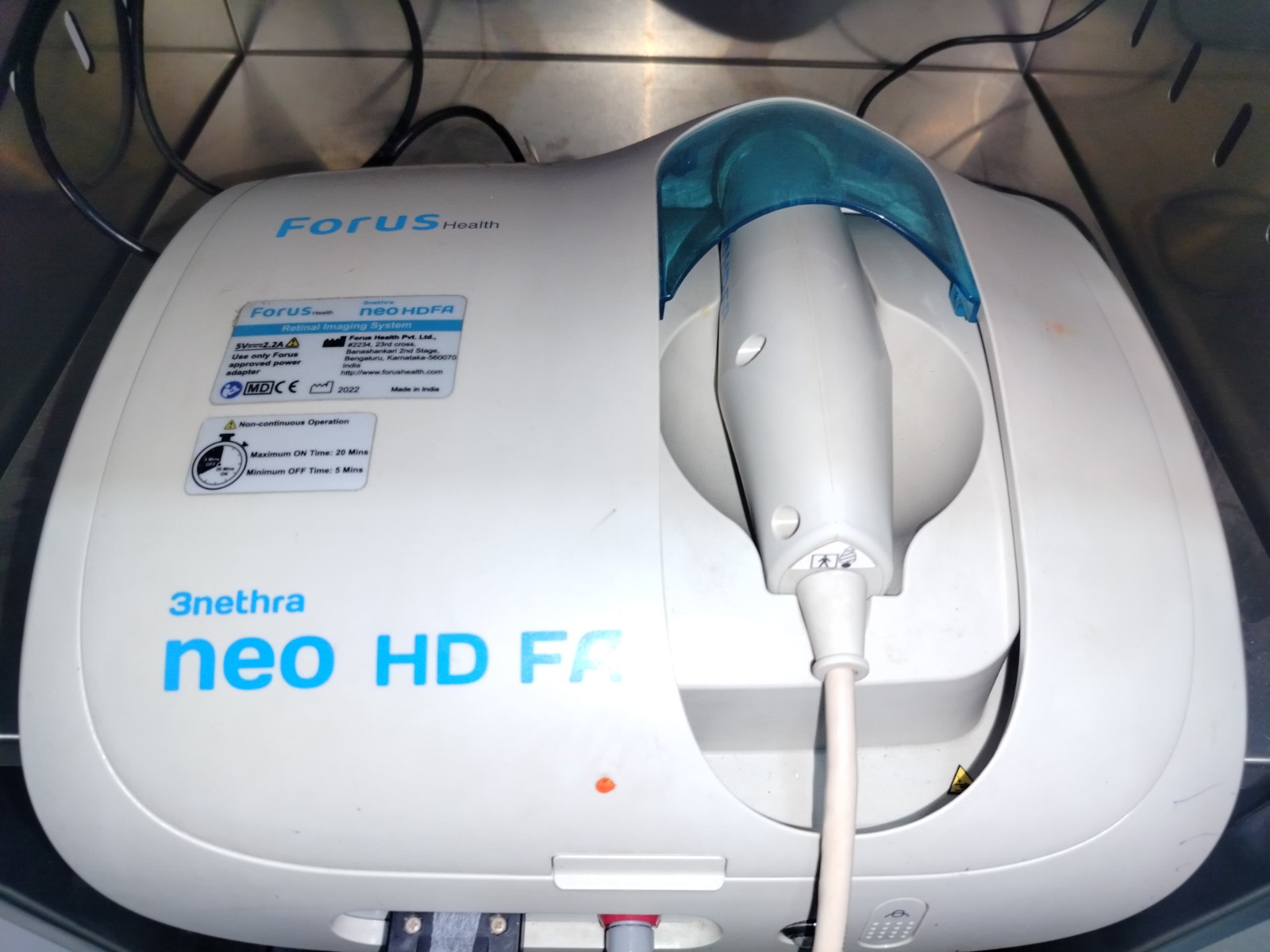
    At Sarakshi Netralaya, our state-of-the-art ROP Clinic in Nagpur offers comprehensive evaluation and treatment for retinal disorders, including hypertensive retinopathy. With advanced retinal imaging and expert ophthalmologists, we ensure early diagnosis and effective follow-up for patients with systemic conditions like hypertension.

    Our focus includes:

    • Routine retinal screening for patients with high blood pressure
    • Digital fundus photography for documentation and follow-up
    • Collaborative care with physicians for systemic hypertension management
    • Specialized care for high-risk patients, including preterm infants (ROP)

    What to Expect in an Eye Check-Up

    • Dilated eye exam
    • Fundus imaging
    • Evaluation of optic nerve and blood vessels
    • Grading of retinal damage
    • Customized treatment plan and monitoring schedule

    Prevention and Care Tips

    • Monitor and control blood pressure regularly
    • Eat a heart-healthy, low-sodium diet
    • Exercise at least 30 minutes a day
    • Avoid smoking and limit alcohol
    • Get routine eye check-ups, especially if you’re over 40 or have a family history of hypertension

    Don’t Wait for Symptoms — Book a Retinal Screening Today

    Hypertension-related eye changes are often silent until it’s too late. Take the proactive step toward protecting your vision. Visit Sarakshi Netralaya’s ROP Clinic in Nagpur for early diagnosis, monitoring, and treatment of hypertensive retinopathy and other retinal conditions.

    Book an Appointment

    [wpforms id=”63″ title=”false”]
  • What’s the difference between open-angle and …

    What’s the difference between open-angle and closed-angle glaucoma?

    Open Angle And Closed Angle Glaucoma Care

    What’s the difference between open-angle and closed-angle glaucoma?

    Glaucoma isn’t one single problem, it’s a family of conditions where pressure inside the eye can hurt the optic nerve over time. Some people feel nothing for years; others get hit with sudden symptoms and need help right away. That’s why doctors talk about two big buckets: open-angle and closed-angle. Same goal (protect the nerve), very different stories. If you just want the short version: one type creeps in quietly, the other barges in loudly. The quieter one is more common. The louder one is rarer but urgent. Let’s unpack both without the jargon overload.

    The slow, sneaky one

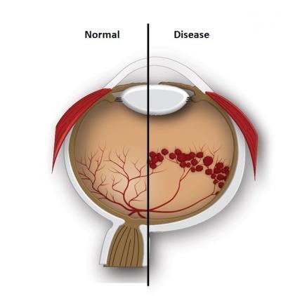
    When folks say open angle glaucoma, they mean the eye’s drainage angle looks open under the microscope, but the tiny filter (trabecular meshwork) doesn’t clear fluid efficiently. Pressure inches up, slowly, sometimes for years, so people don’t notice much at first. Side vision thins out bit by bit; central vision usually stays okay until later. Regular eye checks matter here more than heroics.
    No pain, no redness, vision “seems fine”…until testing says otherwise Gradual loss of peripheral (side) vision The chances of developing glaucoma go up with factors like advancing age, a family history of the condition, thinner-than-average corneas, long-term diabetes, or extended use of steroid medications.

    The sudden, shouty one

    Eye care teams don’t guess, they measure. Pressure checks alone aren’t enough. You’ll likely see a mix of:
    • Gonioscopy (a tiny lens to actually view the angle, open, narrow, blocked?)
    • OCT scans to examine the optic nerve fibers
    • Visual fields to map side-vision gaps
    • Pachymetry (corneal thickness)
    • Sometimes ultrasound/angle imaging if the view is tricky

    What treatment looks like (in real life)

    No one-size-fits-all, but the aim is simple: lower pressure and protect the nerve. Drops are common; some people benefit from laser; some need procedures. For urgent blockage, doctors first relieve the spike, then prevent repeats, often with a tiny laser opening in the iris, and in some cases lens surgery if anatomy is the culprit. If you’ve been googling options and feeling lost, breathe, modern glaucoma eye treatment is a toolbox, not a single hammer. Your plan might start with medicated drops, add laser trabeculoplasty for open angles, or use a laser peripheral iridotomy for angle-closure risk. Your doctor will match the tool to the eye. Over the long haul, many people do well with a mix of routine checks, pressure control, and good follow-through. When your doctor talks timelines, they’re thinking about protecting vision over decades, not just months.

    Quick compare (so it sticks)

    • Open-angle: slow build, usually painless, found on routine exams.
    • Angle-closure: rapid spike, painful/obvious, treat now—not next week.
    • Both can damage the optic nerve if ignored. Both are manageable when caught.

    Everyday questions people actually ask

    • “Can lifestyle help?” — A healthy routine helps overall eye and nerve health.
    • “Will I need surgery?” — Maybe, maybe not. Many people do great on drops or laser alone.
    • “Does screen time cause glaucoma?” — No, but it can cause eye strain/dry eye.

    When to act fast (seriously)

    If you suddenly get eye pain, halos, blurred vision, nausea, and a red eye, don’t wait for morning. That cluster screams angle-closure. Head to urgent eye care or an emergency department that can reach an eye specialist.

    Why sticking with care matters

    Here’s the unglamorous truth: protecting vision is a marathon. Follow-ups catch small changes early. If drops irritate, say so, there are alternatives. If a laser is suggested, ask what it aims to change (flow? angle? both?). Good glaucoma treatment is a partnership; you bring consistency, your doctor brings the right tools at the right time.
    Glaucoma isn’t a life sentence to losing sight. It’s a prompt to get checked, keep pressure in range, and not go missing between appointments. And if you’re worried you’ll forget, set reminders, bring a family member, make it part of your routine. That’s how vision is saved in the real world. If you’re looking for clear guidance and a plan that actually fits your eyes (and your life), our team at Sarakshi Netralaya is happy to help, from first evaluation to long-term care, so you can keep seeing the moments that matter.

    Book an Appointment

    [wpforms id=”63″ title=”false”]
  • Eye donation: All About Donating Eyes

    Eye donation: All About Donating Eyes

    Complete Guide To Voluntary Eye Donation

    Eye donation: All About Donating Eyes

    In a world where medical advancements are solving complex problems, one simple act continues to hold immense power: eye donation. Donating eyes after death can restore vision for those suffering from corneal blindness, a condition that affects millions in India alone. Yet, awareness around eye donation remains limited. This blog breaks down everything one needs to know about eye donation, how it works, who can donate, and why it matters.

    What Is Eye Donation?

    Eye donation refers to the act of donating one’s corneas after death. It is not the entire eye that is transplanted, but the clear front layer called the cornea. This cornea is used to restore sight to individuals who have lost their vision due to corneal damage or disease. Corneal blindness is a major cause of vision loss in India. One person’s decision to donate their eyes can help two others see the world again. The donation is carried out only after death and does not impact funeral rites or appearance.
    Hospitals and eye banks, such as Sarakshi Netralaya, that support eye donation in Nagpur are playing a vital role in making this life-changing act more accessible and better understood.

    Who Can Donate?

    Almost anyone can donate their eyes. The donor’s age, gender, or even blood group usually does not matter. However, a few eligibility factors apply:
    • Anyone above the age of 1
    • Even those with poor eyesight, spectacles, or minor eye issues
    • Individuals with certain chronic conditions (like diabetes or hypertension) can still donate
    • People with communicable diseases like HIV, hepatitis, or rabies are typically excluded

    The Procedure

    The eye donation procedure is simple, respectful, and does not involve any pain or disfigurement. Here’s how it generally works:

    Pledging to Donate

    • Individuals can register with a certified eye bank or hospital.
    • Families can also consent after the individual passes away, even if they haven’t pledged formally.

    After Death

    • It’s important to inform the nearest eye bank as soon as possible after a person passes away.
    • The eyes need to be donated within 4 to 6 hours to keep them suitable for transplant.

    The Retrieval Process

    • A trained technician visits the donor’s home or hospital.
    • The corneas are removed using sterile methods, taking only 20–30 minutes.
    • There is no facial disfigurement.

    Preservation & Transplant

    • The corneas are stored and screened at the eye bank.
    • Once cleared, they are transplanted into a recipient who needs them.

    Why it Matters

    The pros of eye donation extend far beyond restoring vision. It is a gift that lives on, one that has emotional, societal, and psychological benefits. Here are a few reasons why it’s one of the most powerful forms of donation:
    • Restores independence: Sight brings back confidence and autonomy for the recipient.
    • One donor, two lives: Each pair of donated corneas can help two different people see again.
    • Legacy that inspires: Families often report a sense of peace and pride knowing their loved one lives on in another’s vision.
    • Eye donation is completely free.

    Some Common Myths

    Myth: Eye donation disfigures the face.
    Fact: The procedure is non-invasive and respectful. The eyes remain closed, and there’s no noticeable change.

    Myth: Only young people can donate.
    Fact: People of any age can donate as long as the cornea is healthy.

    Myth: You need to die in a hospital.
    Fact: Donations can happen at home too, timely information to the eye bank is key.

    How to Take the First Step

    If you or your family members are interested in pledging your eyes:
    • Connect with an eye bank or hospital like Sarakshi Netralaya.
    • Carry a donor card or mention your wish in official documents.
    • Discuss with your family about your decision.
    Even a conversation can spark a chain reaction that changes lives.
    Eye donation is one of the most meaningful gifts a person can give. It turns the end of one life into a new beginning for someone else. And it all begins with a simple decision. If you ever choose to take that step, we’re here to guide you. Book an Appointment Name Email Phone Department dd-mm-yyyy Appointment Time Send

    Book an Appointment

    [wpforms id=”63″ title=”false”]
  • Foods to Avoid After Laser Eye Surgery for Faster Healing

    Foods to Avoid After Laser Eye Surgery for Faster Healing

    Foods to Avoid After Laser Eye Surgery for Faster Healing

    Eye Surgery Recovery Nutrition

    Avoid These Foods For Faster Eye Surgery Healing

    Laser eye surgery is a life-changing experience that brings newfound clarity and freedom from glasses or contact lenses. But did you know that what you eat after the procedure plays a significant role in how fast and effectively your eyes heal? If you’ve recently undergone laser eye surgery, paying attention to your post-surgery diet is just as important as following your doctor’s care instructions.
    Let’s dive into the foods you should avoid during the recovery phase, so your eyes heal faster, better, and with fewer complications.

    1. Avoid Sugary Snacks and Refined Carbs

    Sweet treats and refined carbs like white bread, pastries, and sugary drinks may taste comforting, but they can cause inflammation and slow down healing. After eye surgery, it’s best to keep blood sugar levels stable, as fluctuating levels can affect the eyes’ ability to recover and increase the risk of infection.
    What to skip
    • Soda, packaged juices
    • Cakes, cookies, and pastries
    • White rice and white bread
    Instead, opt for complex carbs like brown rice or whole grains for steady energy and better healing support.

    2. Say No to Processed and Junk Foods

    Burgers, fries, chips, and heavily processed snacks are loaded with unhealthy fats, salt, and preservatives. These can lead to fluid retention, inflammation, and slower tissue repair, things you definitely want to avoid while focusing on your foods after laser surgery plan.
    Try swapping with
    • Fresh fruits and vegetables
    • Homemade soups and lean proteins
    • Lightly steamed or grilled meals with fewer additives
    Remember, clean eating speeds up clean healing!

    3. Minimize Spicy and Oily Foods

    While a spicy meal might be a staple for many, consuming too much chili or fried items right after surgery can trigger discomfort, acidity, or even cause excessive eye-watering. During this sensitive phase, keep it mild to avoid unnecessary irritation.
    At Sarakshi, equipped with the best laser eye surgeon in Nagpur, we recommend staying off heavily spiced dishes during the first few days of recovery. Your digestive system and your eyes will thank you.
    Foods to avoid here include
    • Deep-fried snacks (samosas, pakoras)
    • Pickles and chutneys loaded with spice
    • Foods with excessive masala or chili powder
    Go for boiled, grilled, or baked foods with minimal seasoning during your recovery phase.

    4. Limit Caffeinated Beverages

    While one cup of coffee or tea is usually fine, going overboard with caffeine can dehydrate your body and your eyes. This can lead to dryness and discomfort, especially when your eyes are trying to recover from the procedure. Whether you’re recovering from eye surgery or elsewhere, hydration is key. Drink plenty of water and switch to herbal teas if you need a warm drink.

    5. Avoid Alcohol Completely During Recovery

    Alcohol has a dehydrating effect and can interfere with medications and post-operative eye drops. It may also delay healing and affect your sleep quality both crucial for recovery.

    Alcohol has a dehydrating effect and can interfere with medications and post-operative eye drops. It may also delay healing and affect your sleep quality both crucial for recovery.

    6. Be Careful with Allergenic or Histamine-Triggering Foods

    Certain foods like shellfish, peanuts, or even fermented products can trigger allergic reactions or histamine release, which can cause inflammation and irritation in the eyes. If you’re following a recovery diet after foods after laser surgery, it’s smart to avoid.
    • Shellfish or sushi
    • Fermented items like kimchi or pickles
    • Strong cheeses like blue cheese or parmesan
    Keep your meals simple, clean, and low on potential allergens during the initial days.

    7. Cut Down on Excessive Salt

    Too much sodium causes your body to retain water and can contribute to puffiness around the eyes. This can cause discomfort and may affect your healing speed.
    Healing after laser eye surgery isn’t just about following medical guidelines it’s also about making small, smart lifestyle choices, starting with your plate. By avoiding these food items, you give your eyes the best chance to recover comfortably, quickly, and without complications.
    At Sarakshi Netralaya, we don’t just offer expert surgical care we guide you through every step of your recovery journey with patient-first advice and continuous support. Still have questions about post-op care or the right diet for your recovery? Talk to our specialists today and experience the most trusted laser eye surgery in Nagpur with a healing plan that works just as clearly as your new vision.

    Book an Appointment

    [wpforms id=”63″ title=”false”]
  • Top 10 Common Workplace Eye Injuries and How to avoid them

    Top 10 Common Workplace Eye Injuries and How to avoid them

    Top 10 Common Workplace Eye Injuries and How to Avoid Them

    Workplace Eye Injury Hazards

    Prevention Of Common Workplace Eye Injuries

    Eyes are among the most sensitive and important parts of the body, and yet they are often exposed to various hazards in the workplace. Whether it’s from chemicals, flying debris, or screen time, workers can face numerous risks that may lead to serious eye injuries. It’s essential to be aware of these common injuries and learn how to avoid them to ensure long-term eye health. In this article, we will explore the top 10 common workplace eye injuries and provide practical tips on how to protect your vision.

    1.Chemical Eye Injuries

    Eye injury caused by chemicals is a common workplace hazard, especially in environments like laboratories, factories, or kitchens. Chemicals can cause immediate damage to the eyes, resulting in irritation, burns, or even permanent vision loss if not treated quickly.
    • Always wear appropriate safety goggles.
    • Ensure that emergency eyewash stations are easily accessible.
    • Follow all safety protocols when using chemicals to minimize exposure.

    2.Foreign Objects in the Eye

    Foreign particles, such as dust, metal shavings, or wood chips can cause irritation, scratches, or even deeper damage to the cornea.
    • Wear safety goggles in environments with flying debris.
    • Avoid touching your eyes with dirty hands.
    • If an object gets into your eye, wash it immediately with clean water.

    3.Blunt Force Trauma

    Blunt force trauma occurs when something strikes the eye or surrounding areas, causing bruising or fractures to the bones around the eye. This type of injury can result in serious damage, including retinal detachment or vision loss. Always wear protective eyewear when working in high-risk environments, such as construction sites or workshops.

    4.UV Rays

    Prolonged exposure to ultraviolet (UV) rays from the sun or artificial lighting can damage the eye, leading to conditions like cataracts or macular degeneration. Use safety glasses or goggles that offer protection for the eyes from harmful UV rays.

    5.Digital Eye Strain

    Many employees spend hours staring at screens, leading to a condition known as digital eye strain. This can cause headaches, dry eyes, blurred vision, and neck pain.
    • Follow the 20-20-20 rule for eyes: Every 20 minutes, take a 20-second break and look at something 20 feet away.
    • Adjust screen brightness and contrast to reduce glare.

    6.Eye Infections

    Eye infections can occur in workplaces with poor hygiene practices or when workers touch their eyes with unwashed hands. These infections can cause redness, discharge, and blurred vision.
    • Wash your hands frequently, especially before touching your face or eyes.
    • Avoid sharing personal items like towels, eye makeup, or contact lenses.
    • Seek medical attention if you experience symptoms of an eye infection.

    7.Laser Eye Injuries

    Laser equipment is widely employed in areas such as manufacturing, research, and healthcare. Laser exposure can cause substantial retinal damage and lasting visual impairment.
    • Always wear protection for the eyes when working with or around lasers.
    • Ensure that laser equipment is properly maintained and operated in a controlled environment.

    8.Exposure to Fumes

    Exposure to smoke or fumes, especially in industrial settings or areas with poor ventilation, can irritate the eyes and lead to long-term damage. Workers in welding, manufacturing, or painting industries are particularly vulnerable.
    • Wear protective goggles to keep smoke and fumes out of your eyes.
    • Work in well-ventilated spaces and utilise proper respiratory protection when needed.

    9.Eye Fatigue

    Eye fatigue, also known as asthenopia, occurs when the eyes are overused, often from prolonged close-up work. Common triggers include excessive screen time, reading, or focusing on tasks that require high precision, such as sewing or assembly work. It can lead to discomfort, strain, and even headaches. Take regular breaks to rest your eyes and reduce strain.

    10.Working in Low-Light

    Low-light conditions, whether indoors or outdoors, can strain the eyes and lead to accidents. Poor visibility can also increase the risk of eye injury from accidents, falls, or collisions.
    • Ensure that work areas are well-lit, particularly if tasks require precision.
    • Use eyewear with built-in lighting, such as headlamps or clip-on lights, if necessary.

    Professional Help

    If you experience any symptoms of an eye injury, such as pain, redness, blurred vision, or irritation, it’s essential to seek immediate medical attention. Delaying treatment can result in permanent damage to your vision. For serious injuries or long-term eye care, visit Sarakshi Netralaya, the Best Eye Hospital in Nagpur, where specialists can diagnose, treat, and offer preventive care for your eyes.
    Eye injuries are common in many workplaces, but they can be prevented with the right precautions. By following simple safety protocols, using protective eyewear, and adopting healthy eye habits like the 20-20-20 rule for eyes, you can significantly reduce your risk of eye injury and ensure long-term eye health.
    If you are in search of an eye clinic in Nagpur, Sarakshi Netralaya is the perfect choice. At Sarakshi, we offer expert care for eye fatigue and other vision-related issues, providing personalized treatment plans to help restore your eye health and comfort. Visit us today to ensure the health of your eyes and take proactive steps toward maintaining your vision for the long term.

    Book an Appointment

    [wpforms id=”63″ title=”false”]
  • Stages of Retinopathy of Prematurity: A Guide for

    Stages of Retinopathy of Prematurity: A Guide for

    Stages of Retinopathy of Prematurity: A Guide for Parents and Caregivers

    Retinopathy Stage Diagnosis

    Understanding Retinopathy Of Prematurity Stages

    The journey of becoming a parent to a premature baby is filled with both hope and challenges. Among the many health concerns that premature infants may face, Retinopathy of Prematurity (ROP) stands out as a critical condition requiring timely diagnosis and intervention. This condition, which primarily affects the eyes, can feel overwhelming, but understanding its stages and treatments can empower parents and caregivers to take the necessary steps for their baby’s well-being.
    At Sarakshi Netralaya, a leading hospital for ROP treatment in Nagpur, we understand the emotional weight of these moments. Our team of skilled specialists is here to guide you through the process, ensuring the best care for your little one.

    What Is Retinopathy of Prematurity?

    Retinopathy of Prematurity occurs when blood vessels in a premature baby’s retina develop improperly. This can cause a variety of vision impairments, ranging from mild to severe, including blindness in extreme cases. The disease usually affects newborns born before 31 weeks’ gestation or weighing less than 1,500 grammes. Early retinopathy of prematurity screening is crucial to identify the condition and start appropriate treatment. With timely intervention, many babies can have a better chance at preserving their vision and living a full, healthy life.

    Understanding the stages

    ROP is categorized into five stages.

    Stage 1: Mild Abnormal Blood Vessel Growth

    In this stage, the blood vessels in the retina begin to develop abnormally. While this stage often resolves on its own without treatment, close monitoring is essential to ensure it does not progress.

    Stage 2: Moderate Abnormal Blood Vessel Growth

    In this stage, the blood vessels in the retina begin to develop abnormally. While this stage often resolves on its own without treatment, close monitoring is essential to ensure it does not progress.

    Stage 3: Severe Abnormal Blood Vessel Growth

    At this point, the blood vessels expand abnormally and begin to pull on the retina. This is a tipping point where therapy is required to avoid irreparable damage.

    Stage 4: Partial Retinal Detachment

    In Stage 4, the retina begins to detach partially due to the abnormal blood vessels tugging on it. Surgery may be required to repair the damage and preserve vision.

    Stage 5: Complete Retinal Detachment

    This is the most advanced stage of ROP, and the retina is completely detached. Vision loss at this stage might be significant, necessitating sophisticated surgical procedures. However, early detection and management can frequently avoid this outcome.

    Causes of Retinopathy of Prematurity

    Understanding the causes of retinopathy of prematurity can help caregivers take preventive measures where possible. The condition is primarily linked to premature birth and low birth weight, as the retinal blood vessels have not had enough time to develop fully.

    Other factors

    • High levels of supplemental oxygen after birth.
    • Difficulty seeing in dim light
    • Infections or complications during birth.
    • Genetic predispositions.

    The Importance of Retinopathy of Prematurity Screening

    Screening is the cornerstone of good ROP management. Babies born preterm should have their first eye test between 4-6 weeks of birth, or earlier if the neonatologist deems it necessary. During the screening, the ophthalmologist examines the baby’s retina for any abnormal growth or changes. This painless procedure, though delicate, is vital in determining the presence and stage of ROP. Timely screenings can prevent complications, giving your baby the best chance at healthy vision.
    Premature Infant Retinopathy
    At Sarakshi Netralaya, we specialize in providing comprehensive eye care, with a dedicated focus on premature infants and ROP. Recognized as a leading hospital for ROP treatment in Nagpur, our team combines state-of-the-art technology with compassionate care.
    Retinopathy of Prematurity (ROP) requires early detection for timely intervention, and at Sarakshi Netralaya, we utilize NEORET, an advanced screening tool designed specifically for newborn eye care. This cutting-edge technology enables precise and non-invasive imaging, allowing our specialists to diagnose ROP at its earliest stages. By integrating NEORET into our neonatal care, we ensure that premature infants receive prompt and effective treatment, significantly reducing the risk of vision impairment. Our commitment to innovation and excellence in pediatric ophthalmology ensures that every newborn gets the best start in life with optimal eye health.

    Book an Appointment

    [wpforms id=”63″ title=”false”]